

Beyond “It Just Happened”: Unpacking the Mechanism of Athletic Injuries
We have all heard the phrase, “I just twisted it,” or “It just gave out.” While these descriptions might convey the immediate sensation of an athletic injury, they barely scratch the surface of how the injury occurred. Understanding the mechanism of an athletic injury – the specific forces, movements, and tissue responses that lead to damage – is paramount for effective treatment, targeted rehabilitation, and, most importantly, future prevention.
Think of it like a detective story. To solve the mystery of an injury, we need to gather clues about the precise sequence of events that led to tissue failure. Modern sports medicine, armed with advanced biomechanical analysis and imaging, allows us to piece together these critical details.
What Exactly is an Injury Mechanism?
The mechanism of an injury describes the how:
- The forces involved: Was it a direct impact, a twisting force, an overstretch, or repeated microtrauma?
- The direction of these forces: From what angle did the force act on the tissue?
- The position of the body/limb: What was the joint position (e.g., knee extended, ankle inverted) when the force was applied?
- The tissue response: Did the tissue stretch, tear, compress, or fracture?
- The speed of the event: Was it a sudden, high-velocity incident or a slower, sustained overload?
By analyzing these elements, we move beyond simply naming the injury (e.g., “ACL tear”) to understanding the event that caused it (e.g., “non-contact deceleration with valgus collapse and internal tibial rotation”).
Common Injury Mechanisms Explained:
Let us look at a few common examples to illustrate different injury mechanisms:
1. Non-Contact Anterior Cruciate Ligament (ACL) Tears: A Cascade of Events
Often seen in sports requiring sudden changes in direction (e.g., soccer, basketball, skiing), non-contact ACL tears are a prime example of a complex injury mechanism. Research consistently points to a combination of factors, rather than a single event (Hewett et al., 2010; Myer et al., 2013).
- The setup: Typically, the athlete is landing from a jump or rapidly decelerating and changing direction.
- Critical movements: The knee often moves into a position of valgus collapse (knee collapsing inward towards the midline), combined with internal rotation of the tibia (shin bone rotating inward relative to the thigh bone), and sometimes hyperextension (knee straightening beyond its normal range).
- The forces: This combined movement places immense rotational and shear forces on the ACL, which ultimately exceeds its tensile strength, leading to a tear.
- Contributing factors: Muscle imbalances (e.g., hamstring weakness relative to quadriceps strength), neuromuscular control deficits, and altered landing mechanics can predispose an athlete to this specific mechanism.
Understanding this mechanism allows for targeted prevention programs focusing on strengthening hip abductors, improving landing mechanics, and enhancing neuromuscular control (Myer et al., 2013).
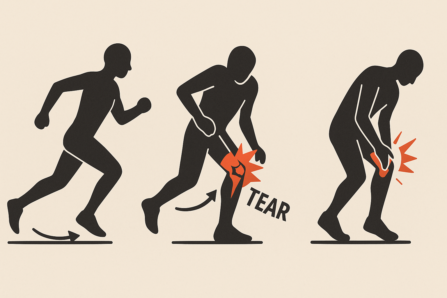
2. Hamstring Strain Injuries: The Overstretch-Contraction Dilemma
Hamstring strains are notoriously common in sprinting and kicking sports. While often attributed to a “pull,” the mechanism is more nuanced. Recent research highlights the role of high-speed running and eccentric muscle contractions (Mendiguchia & Brughelli, 2011; Timmins et al., 2016).
- The setup: Occurs most frequently during the late swing phase of sprinting, just before foot contact, or during rapid changes in direction.
- The critical forces: At this point, the hamstring muscles are rapidly lengthening (eccentric contraction) while simultaneously trying to produce force to decelerate the lower leg and prepare for ground contact.
- The tissue response: If the muscle’s ability to resist this rapid eccentric stretch is exceeded, or if there is a sudden, forceful concentric contraction from an already stretched position, muscle fibers can tear. The biceps femoris long head is often the most affected.
- Contributing factors: Previous hamstring injury, muscle fatigue, inadequate warm-up, and strength imbalances (e.g., between left and right hamstrings, or between hamstrings and quadriceps) increase susceptibility.
This understanding has shifted rehabilitation to prioritize eccentric strengthening and high-speed running drills to prepare the muscle for these specific demands (Timmins et al., 2016).
3. Ankle Sprains: The Inversion Injury
Lateral ankle sprains, involving the ligaments on the outside of the ankle, are one of the most common sports injuries. Their mechanism is typically an inversion injury (Hertel, 2010).
- The setup: Often occurs when landing awkwardly, stepping on an uneven surface, or colliding with another player.
- The critical movement: The foot turns inward (inverts) excessively, while the ankle may also be plantarflexed (pointed downwards).
- The forces: This combination stretches the lateral ankle ligaments (anterior talofibular, calcaneofibular, and posterior talofibular ligaments) beyond their elastic limit, leading to tearing. The anterior talofibular ligament is most commonly affected first.
- Contributing factors: Previous ankle sprains (leading to laxity and reduced proprioception), weak ankle musculature, and improper footwear.
Rehabilitation focuses on restoring balance, proprioception (the sense of joint position), and ankle strength to prevent recurrence (Hertel, 2010).
Why Does Understanding the Mechanism Matter?
- Accurate Diagnosis: Knowing how the injury occurred helps clinicians pinpoint the specific tissues damaged and the extent of the damage.
- Targeted Rehabilitation: Rehabilitation programs can be designed to specifically address the deficiencies or weaknesses that contributed to the injury mechanism. For instance, if valgus collapse leads to an ACL tear, strengthening hip abductors becomes a priority.
- Effective Prevention Strategies: This is perhaps the most powerful application. By identifying common injury mechanisms in a sport or for a specific athlete, coaches and trainers can implement preventative exercises, technique modifications, and training adjustments to mitigate risk (Hewett et al., 2010).
- Improved Return-to-Sport Decisions: Understanding the mechanism helps determine if an athlete has truly re-acquired the strength, control, and movement patterns necessary to safely return to their sport.
Conclusion: From “Oops” to Insight
Moving beyond a superficial understanding of athletic injuries to delve into their underlying mechanisms transforms our approach to sports medicine. It empowers athletes to take a more active role in their prevention, equips coaches with the knowledge to design safer training programs, and provides healthcare professionals with the insights needed for truly effective treatment and rehabilitation. Every injury, no matter how minor, offers a valuable lesson if we are willing to investigate “how” it happened.
References:
- Hertel, J. (2010). Ankle sprains: Mechanisms and prevention. British Journal of Sports Medicine, 44(6), 441-446.
- Hewett, T. E., Myer, G. D., & Ford, K. R. (2010). Anterior cruciate ligament injuries in female athletes: Part 2: A meta-analysis of neuromuscular training programs. American Journal of Sports Medicine, 38(9), 1902-1911.
- Mendiguchia, J., & Brughelli, Y. (2011). A return-to-sport algorithm for acute hamstring injuries. British Journal of Sports Medicine, 45(7), 541-546.
- Myer, G. D., Sugimoto, D., Micheli, L. J., & Hewett, T. E. (2013). Young women’s sports-related ACL injuries: From research to prevention. Current Sports Medicine Reports, 12(5), 291-298.
- Timmins, R. G., Bourne, M. N., Shield, A. J., Williams, M. D., Lorenzen, C., & Opar, D. A. (2016). Short biceps femoris fascicles and hamstring injury in elite sprinters: a prospective cohort study. British Journal of Sports Medicine, 50(23), 1524-1529.
